Agilent FTIR Spectroscopy,
Microscopy and Imaging Solutions
for Biomedical and Biological Applications
Interested in investigating diseased states in animal and human tissue? Use Agilent’s FTIR imaging
microscopy systems to distinguish between cancerous and non-cancerous tissue, develop novel
approaches to assess disease grade and stage, or study the mechanisms of how cells move through
tissues.
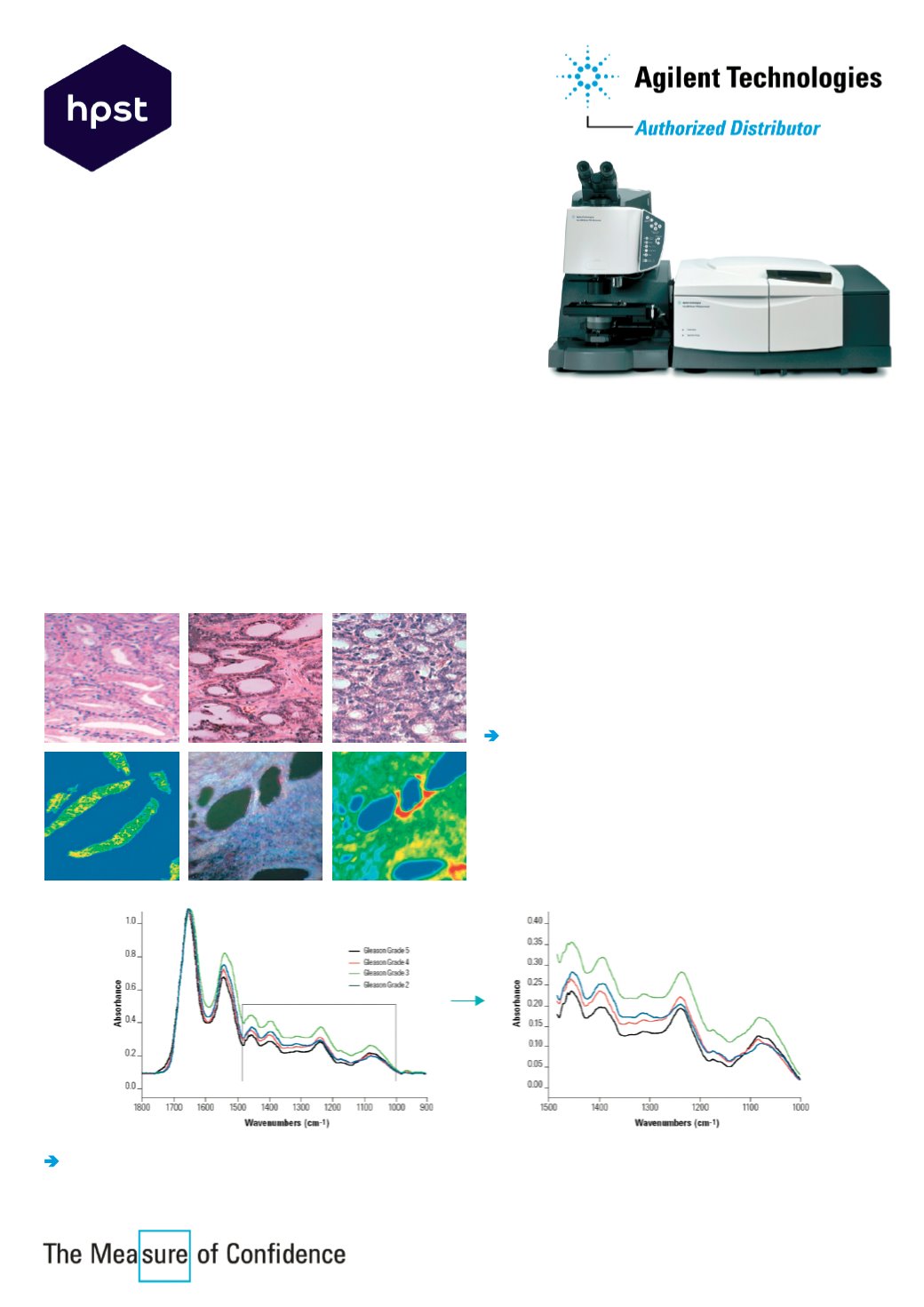
Example: Clinical Diagnosis of Prostate Cancer
Pathologists use an optical microscopy to answer the following questions:
Is it normal or diseased?
Is it BPH or cancer?
If cancer, what grade?
Will it spread?
FTIR imaging – an objective method of analysis:
Representative spectra from different tissue grades
can be used to develop a simple cancer vs non-cancer test and to predict the stage of the disease.
For more information visit
Using optical microscopes
sections of prostate
tissue can be graded according to the Gleason score
based on tissue architecture. But this method suffers
from considerable inter- and intra- observer variability,
i.e. it is a
subjective process
.
An objective method of analysis is needed!
Infrared imaging microscopes
allow for the rapid
screening of large tissue samples. An area of 7x7 mm
(representing 100 tiled images) was recorded with four
pixel aggregation at 8 cm
-1
spectral resolution by co-
adding 16 scans with 11x11 micron spatial resolution.
The entire image took just 40 minutes to collect!
HPST, s.r.o.
Písnická 372/20
142 00 Praha 4
Czech Republic
Tel.: +420 244 001 231
Fax: +420 244 001 235


